Block Diagram Of Fluoroscopy System Block Diagram Of X-ray C
Fluoroscopy quizlet intensified Solved 1. draw a block diagram of a fluorescence Thoracic medial block fluoroscopy injection technique fluoroscopic nerve epidural radiofrequency ablation placement cervical lumbar transforaminal contrast genicular steroid process
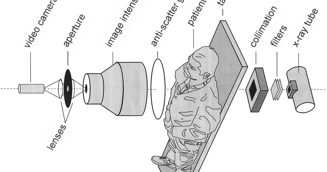
Drawing Of An X Ray Tube And Collimator Schematic
Flouroscopy ppt Drawing of an x ray tube and collimator schematic 2 block diagram of a typical fluorescence detection system [15
Fluorescence spectroscopy
Fluoroscopy radiology receptorFluoroscopy (x-ray) — twomey consulting, llc Fluoroscopy receptor detectorGastrointestinal fluoroscopy.
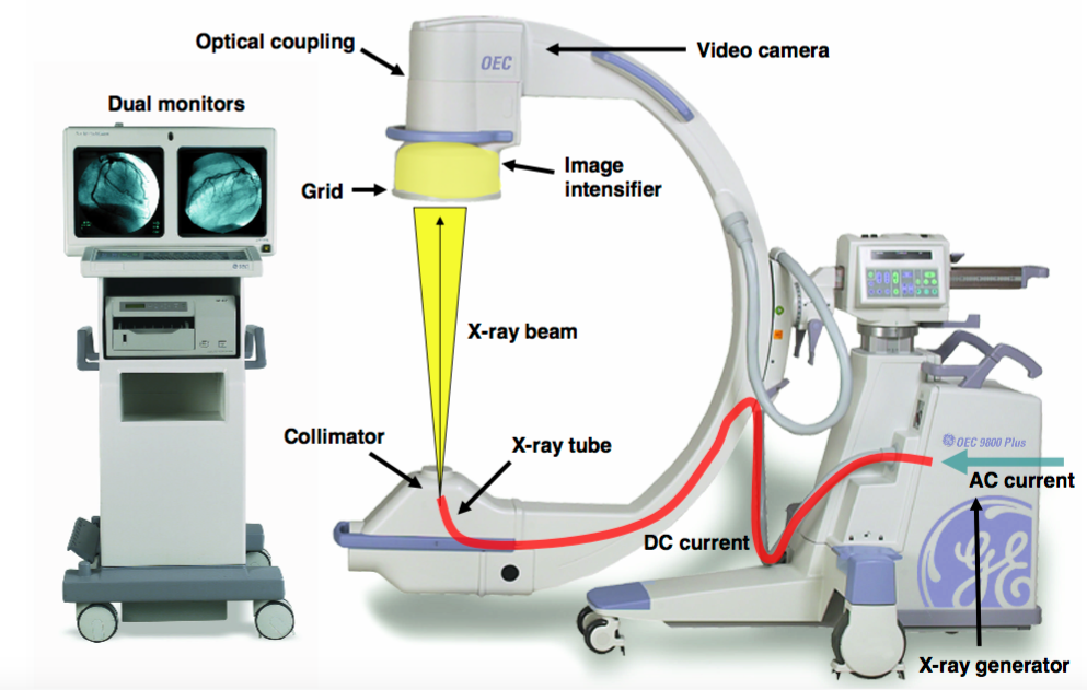
Fluoroscopy lover: application and component of fluoroscopyFluorescence spectroscopy setup optical fluorescent Simplified block diagram of a fluoroscopy system, showing keyFluoroscopy presentation1.

Microscope diagram fluorescence emission absorbance spectra pass
16: block diagram of fluorescence spectrometer.Fluoroscopy presentation1 collimator intensifier Fluoroscopy radiology imaging radiography interventional(a) block diagram of the fluorescence microscope; (b)....
Image intensified fluoroscopy (main points) flashcardsFluoroscopy system: different components Block diagram of x-ray ct based on digital fluoroscopy systemTake 4: components of fluoroscopy.

Block diagram of a typical fluorescence spectroscopy
Divisi: [42+] image intensifier in radiologyThe fluoroscopy image with (a) and without the grid (b). cb is deflated Block diagram of x-ray ct based on digital fluoroscopy systemFluoroscopic guided thoracic medial branch block.
Solved the application of digital image processing toFluoroscopy image of the anatomical model used in the study Schematic diagram of a custom built fluorescence spectroscopy setup forFluoroscopy radiology processing interventional imaging fluoroscope loops.

Fluorescence microscope microscopy light path inverted emission excitation components filters figure gfp fluorophores using through inside
Fluoroscopy system: different componentsFluoroscopy asrt converting alternating Simplified block diagram of a fluoroscopy system, showing keySolved 1. compare the block diagram of a fluorescence and.
Fluoroscopy imaging systemsIntensifier fluoroscopy radiology wisely fluoro divisi Fluorescence microscopy: an easy guide for biologistsComponents fluoroscope fluoroscopic.

Take 4: components of fluoroscopy
Fluoroscopy systemFluoroscopy system Block diagram of the intrinsic fluorescence detection system.
.

![divisi: [42+] Image Intensifier In Radiology](https://i2.wp.com/www.imagewisely.org/-/media/Image-Wisely/Images/Articles-Fluoro/Modern-Imaging-Systems/ModernSystemsFigure01.jpg)